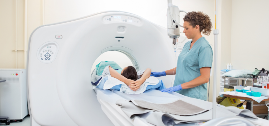
Pelvis Scan

Pelvis Scan
(Ultrasound-Pelvis, Pelvic Ultrasonography, Pelvic Sonography, Pelvic Scan, Lower Abdomen Ultrasound, Gynecologic Ultrasound, Transabdominal Ultrasound, Transvaginal Ultrasound, Endovaginal Ultrasound)
A pelvic ultrasound is a noninvasive demonstrative test that produces pictures that are utilized to evaluate organs and structures inside the female pelvis. A pelvic ultrasound permits snappy perception of the female pelvic organs and structures including the uterus, cervix, vagina, fallopian cylinders and ovaries. Ultrasound utilizes a transducer that conveys ultrasound waves at a recurrence too high to ever be heard. The ultrasound transducer is put on the skin, and the ultrasound waves travel through the body to the organs and structures inside. The sound waves ricochet off the organs like a reverberation and come back to the transducer. The transducer forms the reflected waves, which are then changed over by a PC into a picture of the organs or tissues being analyzed.
The sound waves travel at various rates relying upon the sort of tissue experienced - quickest through bone tissue and slowest through air. The speed at which the sound waves are come back to the transducer, just as the amount of the sound wave returns, is deciphered by the transducer as various kinds of tissue. A ultrasound gel is set on the transducer and the skin to consider smooth development of the transducer over the skin and to dispose of air between the skin and the transducer for the best stable conduction. Another kind of ultrasound is Doppler ultrasound, in some cases called a duplex report, used to show the speed and heading of blood stream in certain pelvic organs. In contrast to a standard ultrasound, some stable waves during the Doppler test are perceptible.
Pelvic ultrasound might be performed utilizing either of 2 strategies:
- Transabdominal (through the stomach area). A transducer is put on the belly utilizing the conductive gel
- Transvaginal (through the vagina). A long, slender transducer is secured with the leading gel and a plastic/latex sheath and is embedded into the vagina
The sort of ultrasound strategy performed relies upon the explanation behind the ultrasound. Just a single technique might be utilized, or the two strategies might be expected to give the data expected to conclusion or treatment. Other related methodology that might be utilized to assess issues of the pelvis incorporate hysteroscopy , colposcopy , and laparoscopy
What are female pelvic organs?
The organs and structures of the female pelvis are:
- Endometrium. The coating of the uterus
- Uterus (otherwise called the belly). The uterus is an empty, pear-molded organ situated in a lady's lower guts, between the bladder and the rectum. It sheds its covering every month during period, except if a prepared egg (ovum) gets embedded and pregnancy follows.
- Ovaries. Two female conceptive organs situated in the pelvis in which egg cells (ova) create and are put away and where the female sex hormones estrogen and progesterone are delivered.
- Cervix. The lower, thin piece of the uterus situated between the bladder and the rectum, shaping a channel that opens into the vagina, which prompts the outside of the body.
- Vagina (otherwise called the birth trench). The way through which liquid drops of the body during menstrual periods. The vagina associates the cervix and the vulva (the outside genitalia).
- Vulva. The outer segment of the female genital organs
What are the purposes behind a pelvic ultrasound?
Pelvic ultrasound might be utilized for estimation and assessment of female pelvic organs. Ultrasound appraisal of the pelvis may incorporate, however isn't restricted to, the accompanying:
- Size, shape, and position of the uterus and ovaries
- Thickness, echogenicity (dimness or softness of the picture identified with the thickness of the tissue), and nearness of liquids or masses in the endometrium, myometrium (uterine muscle tissue), fallopian tubes, or in or close to the bladder
- Length and thickness of the cervix
- Changes fit as a fiddle
- Blood move through pelvic organs
- Vulva. The outer segment of the female genital organs
Pelvic ultrasound can give a lot of data about the size, area, and structure of pelvic masses, however can't give an unequivocal determination of malignant growth or explicit sickness. A pelvic ultrasound might be utilized to analyze and aid the treatment of the accompanying conditions:
Irregularities in the anatomic structure of the uterus, including endometrial conditions
- Fibroid tumors (kind developments), masses, pimples, and different sorts of tumors inside the pelvis
- Nearness and position of an intrauterine preventative gadget (IUD)
- Pelvic provocative illness (PID) and different kinds of aggravation or disease
- Postmenopausal dying
- Checking of ovarian follicle size for fruitlessness assessment
- Yearning of follicle liquid and eggs from ovaries for in vitro treatment
- Ectopic (pregnancy happening outside of the uterus, for the most part in the fallopian tube)
- Observing fetal improvement during pregnancy
- Evaluating certain fetal conditions
Ultrasound may likewise be utilized to help with different systems, for example, endometrial biopsy . Transvaginal ultrasound might be utilized with sonohysterography, a technique where the uterus is loaded up with liquid to widen it for better imaging. There might be different explanations behind your primary care physician to prescribe a pelvic ultrasound.
.png)

